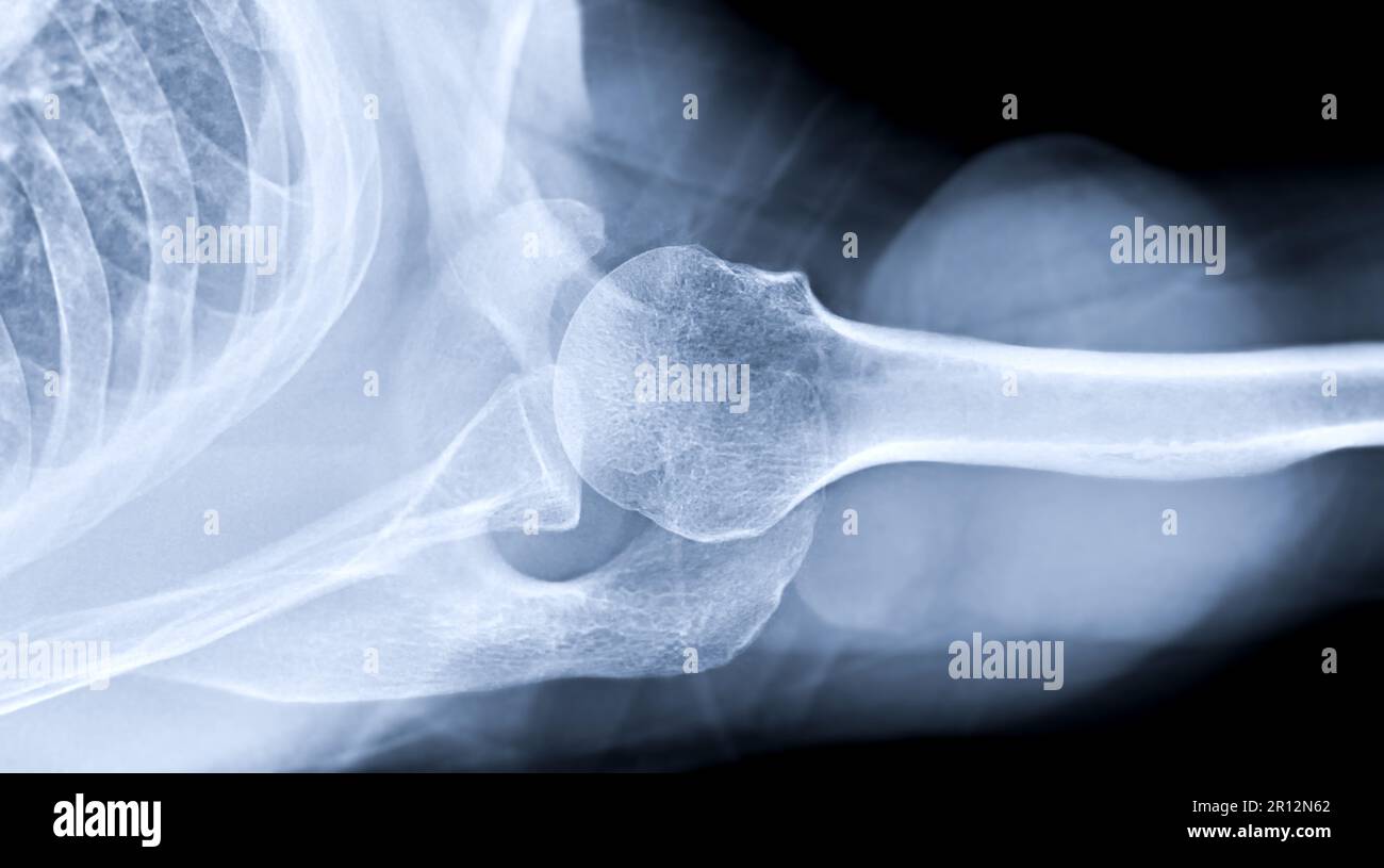
Shoulder Joint Fracture X Ray . diagnosis is made with orthogonal radiographs of the shoulder. Articular surfaces should be parallel. The humeral head should be on the glenoid in any other. A patient with a proximal humerus fracture. Sometimes, additional imaging techniques, such as a. the neer system divides the proximal humerus into four parts and considers not the fracture line, but the displacement as being significant in terms. Front and side pictures show the swelling and bruising down the arm. in this review, we will discuss the mechanisms of injury, key imaging findings, therapeutic options and. Treatment with sling immobilization is indicated for minimally displaced. Magnetic resonance imaging (mri) :.
from www.alamy.com
Magnetic resonance imaging (mri) :. Sometimes, additional imaging techniques, such as a. Treatment with sling immobilization is indicated for minimally displaced. the neer system divides the proximal humerus into four parts and considers not the fracture line, but the displacement as being significant in terms. Front and side pictures show the swelling and bruising down the arm. in this review, we will discuss the mechanisms of injury, key imaging findings, therapeutic options and. The humeral head should be on the glenoid in any other. A patient with a proximal humerus fracture. diagnosis is made with orthogonal radiographs of the shoulder. Articular surfaces should be parallel.
Xray Shoulder joint shoulder transaxillary view for diagnosis fracture of shoulder joint Stock
Shoulder Joint Fracture X Ray Sometimes, additional imaging techniques, such as a. The humeral head should be on the glenoid in any other. diagnosis is made with orthogonal radiographs of the shoulder. Sometimes, additional imaging techniques, such as a. in this review, we will discuss the mechanisms of injury, key imaging findings, therapeutic options and. Magnetic resonance imaging (mri) :. Treatment with sling immobilization is indicated for minimally displaced. Front and side pictures show the swelling and bruising down the arm. Articular surfaces should be parallel. the neer system divides the proximal humerus into four parts and considers not the fracture line, but the displacement as being significant in terms. A patient with a proximal humerus fracture.
From www.dreamstime.com
Xray Shoulder Joint Shoulder Transaxillary View for Diagnosis Fracture of Shoulder Joint Stock Shoulder Joint Fracture X Ray The humeral head should be on the glenoid in any other. A patient with a proximal humerus fracture. diagnosis is made with orthogonal radiographs of the shoulder. Magnetic resonance imaging (mri) :. in this review, we will discuss the mechanisms of injury, key imaging findings, therapeutic options and. Articular surfaces should be parallel. Sometimes, additional imaging techniques, such. Shoulder Joint Fracture X Ray.
From www.alamy.com
Xray Shoulder joint shoulder transaxillary view for diagnosis fracture of shoulder joint Stock Shoulder Joint Fracture X Ray Front and side pictures show the swelling and bruising down the arm. Articular surfaces should be parallel. the neer system divides the proximal humerus into four parts and considers not the fracture line, but the displacement as being significant in terms. in this review, we will discuss the mechanisms of injury, key imaging findings, therapeutic options and. . Shoulder Joint Fracture X Ray.
From buyxraysonline.com
ANTERIOR SHOULDER DISLOCATION WITH FRACTURE Shoulder Joint Fracture X Ray Sometimes, additional imaging techniques, such as a. the neer system divides the proximal humerus into four parts and considers not the fracture line, but the displacement as being significant in terms. diagnosis is made with orthogonal radiographs of the shoulder. The humeral head should be on the glenoid in any other. A patient with a proximal humerus fracture.. Shoulder Joint Fracture X Ray.
From www.alamy.com
Xray of both Shoulder joint AP view for diagnosis fracture of shoulder joint Stock Photo Alamy Shoulder Joint Fracture X Ray Treatment with sling immobilization is indicated for minimally displaced. Magnetic resonance imaging (mri) :. Sometimes, additional imaging techniques, such as a. The humeral head should be on the glenoid in any other. diagnosis is made with orthogonal radiographs of the shoulder. Articular surfaces should be parallel. Front and side pictures show the swelling and bruising down the arm. . Shoulder Joint Fracture X Ray.
From buyxraysonline.com
ACROMIOCLAVICULAR JOINT INJURY Shoulder Joint Fracture X Ray Front and side pictures show the swelling and bruising down the arm. diagnosis is made with orthogonal radiographs of the shoulder. The humeral head should be on the glenoid in any other. in this review, we will discuss the mechanisms of injury, key imaging findings, therapeutic options and. the neer system divides the proximal humerus into four. Shoulder Joint Fracture X Ray.
From www.shutterstock.com
Стоковая фотография 1588507039 Xray Shoulder Joint Fracture Humerus Medical Shutterstock Shoulder Joint Fracture X Ray Articular surfaces should be parallel. diagnosis is made with orthogonal radiographs of the shoulder. Front and side pictures show the swelling and bruising down the arm. Sometimes, additional imaging techniques, such as a. The humeral head should be on the glenoid in any other. the neer system divides the proximal humerus into four parts and considers not the. Shoulder Joint Fracture X Ray.
From www.researchgate.net
Anteroposterior radiograph of the left shoulder shows a pathological... Download Scientific Shoulder Joint Fracture X Ray Front and side pictures show the swelling and bruising down the arm. diagnosis is made with orthogonal radiographs of the shoulder. in this review, we will discuss the mechanisms of injury, key imaging findings, therapeutic options and. Treatment with sling immobilization is indicated for minimally displaced. Articular surfaces should be parallel. A patient with a proximal humerus fracture.. Shoulder Joint Fracture X Ray.
From www.alamy.com
Xray of Shoulder joint showing fracture of humerus bone Stock Photo Alamy Shoulder Joint Fracture X Ray Articular surfaces should be parallel. Sometimes, additional imaging techniques, such as a. The humeral head should be on the glenoid in any other. in this review, we will discuss the mechanisms of injury, key imaging findings, therapeutic options and. Magnetic resonance imaging (mri) :. Front and side pictures show the swelling and bruising down the arm. A patient with. Shoulder Joint Fracture X Ray.
From www.dreamstime.com
Xray Shoulder Fracture Posterior Half of Glenoid with Posterior Dislocation of the Bone Shoulder Joint Fracture X Ray Treatment with sling immobilization is indicated for minimally displaced. Sometimes, additional imaging techniques, such as a. The humeral head should be on the glenoid in any other. diagnosis is made with orthogonal radiographs of the shoulder. the neer system divides the proximal humerus into four parts and considers not the fracture line, but the displacement as being significant. Shoulder Joint Fracture X Ray.
From 4.bp.blogspot.com
Xray.Shoulder.Fracture (image) Shoulder Joint Fracture X Ray the neer system divides the proximal humerus into four parts and considers not the fracture line, but the displacement as being significant in terms. A patient with a proximal humerus fracture. in this review, we will discuss the mechanisms of injury, key imaging findings, therapeutic options and. Treatment with sling immobilization is indicated for minimally displaced. Articular surfaces. Shoulder Joint Fracture X Ray.
From www.alamy.com
Xray Shoulder joint shoulder front view for diagnosis fracture of shoulder joint Stock Photo Shoulder Joint Fracture X Ray A patient with a proximal humerus fracture. the neer system divides the proximal humerus into four parts and considers not the fracture line, but the displacement as being significant in terms. The humeral head should be on the glenoid in any other. in this review, we will discuss the mechanisms of injury, key imaging findings, therapeutic options and.. Shoulder Joint Fracture X Ray.
From www.cureus.com
Cureus Bilateral Simultaneous Asymmetrical Anterior Shoulder Dislocation With a Fracture Shoulder Joint Fracture X Ray The humeral head should be on the glenoid in any other. diagnosis is made with orthogonal radiographs of the shoulder. A patient with a proximal humerus fracture. Articular surfaces should be parallel. Front and side pictures show the swelling and bruising down the arm. Treatment with sling immobilization is indicated for minimally displaced. Magnetic resonance imaging (mri) :. . Shoulder Joint Fracture X Ray.
From www.cureus.com
Cureus Bilateral Simultaneous Asymmetrical Anterior Shoulder Dislocation With a Fracture Shoulder Joint Fracture X Ray A patient with a proximal humerus fracture. Sometimes, additional imaging techniques, such as a. The humeral head should be on the glenoid in any other. in this review, we will discuss the mechanisms of injury, key imaging findings, therapeutic options and. diagnosis is made with orthogonal radiographs of the shoulder. Magnetic resonance imaging (mri) :. Articular surfaces should. Shoulder Joint Fracture X Ray.
From sydneyshoulderunit.com.au
Shoulder Fracture with Dislocation Medical Case Study Sydney Shoulder Unit Shoulder Joint Fracture X Ray in this review, we will discuss the mechanisms of injury, key imaging findings, therapeutic options and. A patient with a proximal humerus fracture. Magnetic resonance imaging (mri) :. Sometimes, additional imaging techniques, such as a. the neer system divides the proximal humerus into four parts and considers not the fracture line, but the displacement as being significant in. Shoulder Joint Fracture X Ray.
From stock.adobe.com
Xray Shoulder joint shoulder transaxillary view for diagnosis fracture of shoulder joint. Stock Shoulder Joint Fracture X Ray Sometimes, additional imaging techniques, such as a. Front and side pictures show the swelling and bruising down the arm. The humeral head should be on the glenoid in any other. the neer system divides the proximal humerus into four parts and considers not the fracture line, but the displacement as being significant in terms. in this review, we. Shoulder Joint Fracture X Ray.
From sanjaygarude.com
Shoulder Fractures Dr. Sanjay Garude Shoulder Joint Fracture X Ray the neer system divides the proximal humerus into four parts and considers not the fracture line, but the displacement as being significant in terms. Magnetic resonance imaging (mri) :. in this review, we will discuss the mechanisms of injury, key imaging findings, therapeutic options and. A patient with a proximal humerus fracture. diagnosis is made with orthogonal. Shoulder Joint Fracture X Ray.
From www.alamy.com
Xray Shoulder joint shoulder transaxillary view for diagnosis fracture of shoulder joint Stock Shoulder Joint Fracture X Ray Front and side pictures show the swelling and bruising down the arm. in this review, we will discuss the mechanisms of injury, key imaging findings, therapeutic options and. Treatment with sling immobilization is indicated for minimally displaced. diagnosis is made with orthogonal radiographs of the shoulder. Sometimes, additional imaging techniques, such as a. The humeral head should be. Shoulder Joint Fracture X Ray.
From www.pinterest.com
Rt shoulder pathologic fracture metastases Radiology, Medical dental, Pathology Shoulder Joint Fracture X Ray Articular surfaces should be parallel. Sometimes, additional imaging techniques, such as a. in this review, we will discuss the mechanisms of injury, key imaging findings, therapeutic options and. diagnosis is made with orthogonal radiographs of the shoulder. The humeral head should be on the glenoid in any other. A patient with a proximal humerus fracture. Treatment with sling. Shoulder Joint Fracture X Ray.